Hey there! This is your friendly neighborhood REEF intern, coming to you once more from sunny Key Largo. The internship is a great way to learn new skills and gain experience. While there is a lot of new learning, there are also opportunities to apply knowledge and abilities that we already possess. While many of many colleagues throughout my recent marine science career have backgrounds in marine biology and oceanography, I come from a bit of a different path. My background is in Earth science and Geoinformatics, a fancy word to describe the use of technology such as remote sensing, mapping, and GIS to address specific tasks or problems. My specialty is in structure from motion photogrammetry, which is the method in which a series of images that cover the entire area of an object or plot of land can be used to create a 3 dimensional rendering for data collection and analysis. So far I have used the technology quantify oyster biomass on artificial reefs off the Delmarva Peninsula, as well as map coral reefs on the island of Curaçao. This summer, REEF gave me the chance to add a few more feathers to my cap, and use my special skill set.
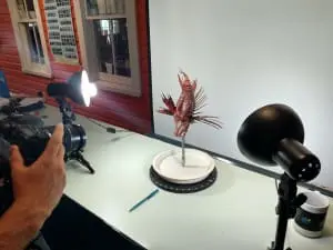
The first opportunity was in creating a 3D lionfish model for education purposes. The idea is that a model is created of the whole lionfish, then one of a fish that had been descaled to expose muscle tissue, and finally one of a fish that had been filleted, revealing the underlying skeletal structure. By creating the series of lionfish models, it would be possible to perform on online dissection of the fish, a handy tool for education. I came into the project rather confident. I had just modeled oyster colonies, full of little nooks and crannies that are nightmares to capture. It is just a fish, how hard can it be? Boy was I wrong. While the body was simple enough, two areas emerged as areas of trouble and frustration. The first area was the long, thin spines found on the fish’s back. They were just thin enough that parts of the spines would not appear on the final model. The second area was the anal and rear fins. While they do have some pattering on them, their mostly translucent nature brought about difficulties in recognition by the modeling software. Over the course of 2 and a half months, 12 trials and fish models, various solutions were discovered to tackle the problems.


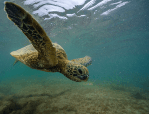
The second opportunity was to continue my main passion, applying the technology to the mapping of corals and coral reefs for monitoring reef health and heavily impacted areas. My introduction to the world of coral imaging and rendering began as an oceanography project. I’ve since stepped up from the tiny GoPro sensor with 5 megapixel stills, to a nicer point and shoot with RAW capability. Thanks to REEF’s location, situated right by the only coral reef in the continental US, I had the equipment, the knowhow, and the opportunity to continue with my personal project. Using some of my allotted half-days of diving, I went out on the reefs and tried various different methods to see which worked best. I also attempted to model different corals to see what techniques would work best for each corresponding structure type. I worked on imaging and modeling Acropora palmate (staghorn coral), a big change from the Montastrea (Boulder) , Diploria (Brain), and Dendrogyra (Pillar) species that I was used to working with. My success was varied, however my methodology and experience has vastly improved and increased.

And that is just another day as a REEF intern. The diversity of what one gets to do as an intern is unrivaled. Never a dull day.